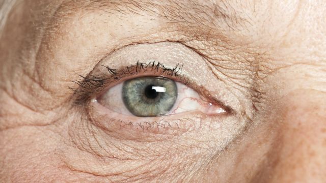
According to Syed Jamal, retinal health can potentially serve as a predictive factor for brain function (Sherman Smith/Kansas Reflector).
The eye, often described as a window to the soul, can also provide valuable insights into the workings of the brain.
The eye has a strong connection to the brain. The retina, located at the back of the eye, acts as a film where images are formed before being transmitted to the brain through the optic nerve.
Maintaining eye health is crucial for older adults, particularly those above the age of 65. Recent studies have highlighted a potential link between eye health and the risk of developing Alzheimer’s and dementia. Therefore, it is important to raise awareness and prioritize the well-being of our eyes as we age.
Recent studies suggest that the health of the retina may serve as a predictive measure for brain function. These studies indicate that retinal health is linked to our ability to remember names and events, appreciate fragrances, and recognize the faces of our loved ones.
A study conducted over a span of 14 years examined retinal and brain tissue samples from 86 individuals. The participants included normal donors, donors experiencing mild cognitive declines, and those in the late stages of Alzheimer’s disease. The study revealed a notable increase in beta amyloid levels and a substantial decrease in microglial cells in individuals with cognitive issues. Microglia, which play a crucial role in repairing injuries and maintaining the neuronal network, were found to be significantly affected.
Beta-amyloid, a byproduct of APP, is a membrane protein that undergoes enzymatic breakdown into various products. It serves as a marker for Alzheimer’s disease and early cognitive decline.
Inflammation in the far region of the retina may also indicate a potential decline in cognitive function.
A recent study introduces an innovative method known as Optical Coherence Tomography (OCT) to uncover insights about retinal thickness and forecast the likelihood of developing mild cognitive impairment and Alzheimer’s disease.
OCT utilizes an infrared beam of light to target specific tissue, merging it with a reference beam. By analyzing the resulting interference patterns, scans are generated, revealing the scattering properties of the tissues in the beam’s trajectory. The light beam is then systematically moved along the tissue, capturing a series of scans. This innovative technology allows for the measurement of blood flow to the retinal vessels, enabling the detection of any decreases in blood flow to the optic papilla or disc.
When using an ophthalmoscope, we can observe the optic disc as a circular, yellowish-pink structure. This instrumental tool allows us to examine the retina, optic nerve, and the blood vessels that provide nourishment to the retina. Interestingly, the optic disc is often referred to as the blind spot, owing to its absence of photoreceptors. This concept of blind spots is commonly associated with the cautionary reminder to check our blind spots while driving and changing lanes on the road.
The optic disc serves as the gateway for the optic nerve and the blood vessels that nourish the eye. Glaucoma, a condition characterized by elevated fluid pressure, often targets the optic disc. The retinal artery, a branch of the internal carotid artery, and its tributaries play a crucial role in supplying blood to the retina and other components of the eye.
A meta-analysis of 11 studies revealed that individuals with Alzheimer’s disease experience a decrease in retinal thickness. Surprisingly, this abnormality extends throughout the entire retinal layer, indicating that it is not solely attributed to the natural aging process. While the exact biological mechanisms behind the degenerative processes that cause structural changes in the brains of Alzheimer’s patients are still being explored, some scientists speculate that these same processes also impact the neural layers of the retina.
The study had certain limitations. Firstly, the sample size was not large enough to generalize the results. Additionally, the use of seven different types of OCT instruments may have affected the reliability of the findings. It is important to note that when different instruments are used, the results may vary. Moreover, the involvement of seven individuals using different instruments could introduce bias into the results.
These findings provide compelling evidence to support further research and serve as a reminder for primary care doctors to inquire about the eye health of patients over the age of 45. If any issues are suspected, healthcare providers can promptly refer patients to an ophthalmologist for further evaluation and treatment.
Exciting times are upon the field of eye research as the convergence of artificial intelligence, big data, and advanced imaging techniques has given rise to a new discipline called oculomics. This burgeoning field aims to identify ophthalmic biomarkers that can provide insights into other diseases within the body. However, delving deeper into oculomics is a topic for another discussion.
Syed Jamal is a college-level chemistry, biology, and anatomy/physiology professor who conducts research on phytoremediation and cancer biology. The Kansas Reflector aims to give a platform to individuals who are impacted by public policies or marginalized in public discussions. For more details, including guidelines on submitting your own commentary, please visit their website.
Eye health could potentially play a crucial role in detecting Alzheimer’s and dementia at an early stage, according to recent research. This new study sheds light on the vital connection between eye health and cognitive decline. The findings emphasize the importance of regular eye examinations as a potential tool for early diagnosis and intervention. It is crucial to recognize the significance of eye health in identifying and addressing cognitive disorders. By prioritizing eye health, it may be possible to detect Alzheimer’s and dementia earlier, facilitating timely treatment and support for those affected.

